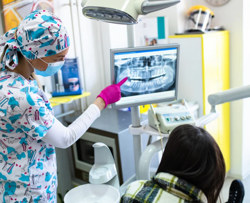
Digital X-Rays
Digital radiographs, commonly known as x-rays, are essential tools for diagnosing and assessing dental health. At our practice, we use modern digital x-rays to enhance both the convenience and efficiency of your treatment.
In the past, dental x-rays were captured using film, similar to analog photography. With the rise of digital imaging, computerized radiography has become the industry standard, reducing radiation exposure by up to 90% compared to traditional film x-rays. Digital x-rays use a small electronic sensor instead of silver-oxide film, eliminating the need for environmentally harmful development chemicals.

What Is A Digital X-Ray?
Radiographs, commonly referred to as X-rays, have been integral to preventive dental care for many years. X-rays are a type of invisible electromagnetic radiation. When these rays were first discovered, their nature was unknown, leading to their designation as X-rays—a name that has endured.
X-rays can penetrate the soft tissues of the face and mouth (such as lips and cheeks) but are absorbed by the hard tissues of teeth and bone. This characteristic enables dentists to detect oral health issues not easily visible externally. X-rays are primarily used to identify cavities, but they also help in examining tooth roots, evaluating the bone health around teeth, assessing potential periodontal (gum) disease, analyzing tooth and jaw alignment, and monitoring development in younger patients.
Type of Dental Digital X-Rays
Among the various types of dental X-rays, bite-wing X-rays are the most commonly used. Named after the wing-like shape of the films once used, these X-rays are taken while you sit in the dental chair and capture images of multiple teeth, including their roots. A dental team member will position a sensor in your mouth and ask you to bite down while aiming a tube-shaped device at your face. This device, known as the X-ray emitter, projects X-rays through your tissues onto the sensor. The process involves no light or heat, and typically, there is no discomfort associated with taking dental X-rays.
Bitewing X-Rays
Capture the upper and lower teeth in a single view, often used to detect cavities between teeth and monitor bone levels.
Periapical X-Rays
Focus on the entire tooth, from the crown to the root, and the surrounding bone, helping to identify issues like abscesses or bone loss.
Panoramic X-Rays
Provide a broad view of the entire mouth, including the teeth, jaws, nasal area, and sinuses. They are useful for planning orthodontic treatments or assessing impacted teeth.
Occlusal X-Rays
Show the roof or floor of the mouth and help detect abnormalities, such as cysts or impacted teeth.
Cone Beam CT (CBCT) Scans
Offer 3D images of the teeth, soft tissues, nerve pathways, and bone, providing detailed information for complex cases like implants or reconstructive surgery.
The Advantage of Modern Digital X-Rays
A major advantage of modern digital X-rays over traditional film is the immediate availability of images. With digital X-rays, the image of your tooth can be quickly transmitted to a monitor in the treatment room, allowing the dentist to view your teeth and surrounding structures while you’re still in the chair. This instant access enables the dentist to evaluate your oral health and identify potential issues right away. The dentist can easily highlight problem areas, helping you understand your oral health condition as they explain it. Additionally, digital files can be effortlessly shared with other dental professionals who may be involved in your care.
Contact Us Today
to schedule an initial consultation & exam.
Your consultation will include an examination of everything from your teeth, gums and soft tissues to the shape and condition of your bite. Generally, we want to see how your whole mouth looks and functions. Before we plan your treatment we want to know everything about the health and aesthetic of your smile, and, most importantly, what you want to achieve so we can help you get there.
